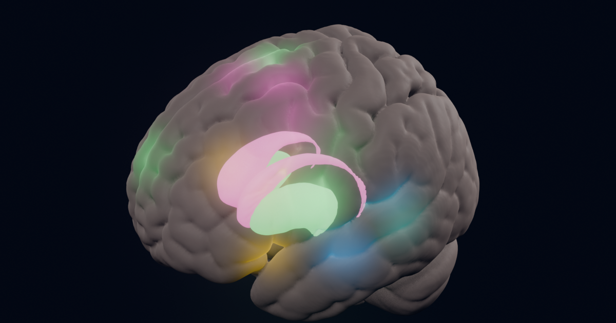
This is a look inside the interpreting brain. A simultaneous interpreter listens to one language and speaks another at the same time, a couple of seconds behind the speaker. In a hospital, a courtroom or a meeting, the same interpreter often works both ways: a doctor’s English into Spanish, then the patient’s Spanish back into English. Brain-imaging studies have measured which parts of the brain work harder while people do this. The animations below show those regions on a real brain, at the places the studies report.
What “lighting up” means
The main study here, Hervais-Adelman et al. 2015, scanned 50 multilingual people with functional MRI (fMRI) while they listened to sentences in English or French, repeated them (“shadowing”), or interpreted them into their most fluent language. fMRI shows where blood flow rises, a sign of harder-working brain tissue, averaged over seconds and over the group. Comparing interpreting with repeating the same sentences shows what the translation itself adds, since both involve hearing and speaking.
So the animation is a map, not a film of thoughts: fMRI cannot show the order in which these regions act within a second. We show them one at a time, slowed down, to explain what each does. In a real interpreter they all work together, continuously.
The interpreting brain, step by step
- Hearing. The auditory cortex along both temporal lobes follows the speaker. In the study its activity rose with the time spent listening and speaking at once (2.1 seconds of overlap per sentence on average).
- Meaning and the words in the other language. The left inferior frontal gyrus, the front part of Broca’s area (pars triangularis and pars orbitalis), and the left anterior insula: regions linked to finding words and meanings.
- Keeping the right language in charge. The caudate nucleus, deep in the brain, with the pre-supplementary motor area and the anterior cingulate near the top. The authors describe the caudate as taking part in “the overarching selection and control of the lexico-semantic system”: keeping the target language in use and the other one in check. This is the same network used for self-control in tasks that have nothing to do with language.
- Speaking while still listening. The motor cortex, the supplementary motor area and the cerebellum produce speech. The putamen, beside the caudate, worked harder the longer the interpreter had to speak and listen at once: the authors link it to “ongoing control of language output”.
Two ways: the direction changes the work
Interpreters usually work into their native language, which is considered the easier direction, and the study above did the same. Working both ways adds the other direction. Rinne et al. 2000 measured professional interpreters with PET while they interpreted between Finnish and English. Into their native Finnish, left frontal activity increased. Into English, their second language, activity increased over “much more extensive” left frontal and temporal areas. That study’s abstract gives no coordinates, so the wider spread on the right of the animation is an illustration of “more”, placed around the same left-side regions.
A two-way interpreter also switches direction at every turn of the conversation, which calls on the control network (in magenta) again and again.
Practice makes it more automatic
Hervais-Adelman, Moser-Mercer & Golestani 2015 scanned students at the start and the end of a master’s course in conference interpreting, and people in other courses. After training, the interpreters’ right caudate nucleus was recruited less while interpreting. The authors take this as the task becoming more automatic, needing less control.
How the animations were made
The brain is the MNI ICBM152 2009c template, an average of 152 adult brains from the Montreal Neurological Institute (McGill University), the same reference brain the studies report their coordinates in. The caudate and putamen come from the CIT168 atlas (Pauli, Nili & Tyszka, 2018). Each glow sits at a peak listed in the study’s tables, its strength scaled by the peak’s statistical score. We rendered it in Blender.
More articles like this one are in our Research section.
Sources
- Hervais-Adelman A, Moser-Mercer B, Michel CM, Golestani N (2015). fMRI of simultaneous interpretation reveals the neural basis of extreme language control. Cerebral Cortex 25(12):4727–4739. doi:10.1093/cercor/bhu158 (free: University of Geneva)
- Hervais-Adelman A, Moser-Mercer B, Golestani N (2015). Brain functional plasticity associated with the emergence of expertise in extreme language control. NeuroImage 114:264–274. doi:10.1016/j.neuroimage.2015.03.072
- Rinne JO, Tommola J, Laine M, et al. (2000). The translating brain: cerebral activation patterns during simultaneous interpreting. Neuroscience Letters 294(2):85–88. doi:10.1016/S0304-3940(00)01540-8
- Fonov V, Evans AC, Botteron K, et al. (2011). Unbiased average age-appropriate atlases for pediatric studies. NeuroImage 54(1):313–327 (MNI ICBM152 2009c; Copyright © 1993–2004 Louis Collins, McConnell Brain Imaging Centre, Montreal Neurological Institute, McGill University)
- Pauli WM, Nili AN, Tyszka JM (2018). A high-resolution probabilistic in vivo atlas of human subcortical brain nuclei. Scientific Data 5:180063. doi:10.1038/sdata.2018.63 (CC BY 4.0)